Case report: Effortless high-quality veterinary dental imaging with Planmed Verity®
Blog post
1 August 2019

When you need detailed information about an animal’s mouth and skull, cone beam computed tomography (CBCT) imaging is the best way to see it all. CBCT imaging provides highly accurate and detailed images for assessment and treatment planning in only a few minutes. Planmeca Group offers the mobility and ease of accessibility of CBCT imaging for veterinary dentists for use on animals of all shapes and sizes - even a jaguar with a damaged canine tooth.
Inka is a five-year-old jaguar in residence at the Elmwood Park Zoo in Pennsylvania, USA. For jaguars to reach their full life expectancy of up to 23 years in captivity, the preservation of their health is essential for zoos to protect this near threatened species.
Jaguars use their bite to suffocate and crush their prey and ingest bones and have the strongest jaw of any feline. The preservation of their dental health is especially vital. Inka was treated with pulp therapy a few years ago when her canine tooth was fractured. Unfortunately, Inka suffered a stroke-like event after a previous surgical procedure but was able to recover.
Two years later, in 2018, the zoo staff started to suspect that the treated canine or other teeth might be the source of pain for Inka. The zoo wanted to take no unnecessary risks with potential complications and decided to look for alternative ways to determine a diagnosis and treatment plan.
Planmeca sales representative Jeff Scharff took a tactical approach and provided veterinary dental specialist Dr John Lewis the use of a Planmed Verity® CBCT unit. The agile and low-profile CBCT veterinary scanner was set cage-side at the zoo – right where it was needed to lessen the transfer of Inka. Planmed Verity’s integrated video displayed a view of Inka, adding confidence in proper patient positioning. The Verity scanned the complete skull in under 20 seconds. In an additional 60 seconds, the high-resolution 3D volumetric images were ready for review.

Preparing Inka for the CBCT scan.

Inka positioned in the Planmed Verity® scanner.
CBCT scanning is a non-invasive method of veterinary dental imaging and especially suitable for cases where accurate information of the full dentition and skull is required. The expanse of detail can be essential for a precise diagnosis and treatment plan, especially if multiple teeth are causing symptoms. A CBCT scans the whole mouth and skull in a fraction of the time compared to taking numerous intraoral scans of the teeth and with less detail of the surrounding anatomy. Radiation exposure and procedure time the time under anaesthesia are reduced, which in turn, minimises post-operative complications.
CBCT offers superior diagnostic yield of pathology for assessment with highly accurate 3D scans. CBCT is particularly suitable for use on small to medium-sized brachycephalic animals due to the increased level of detail available compared to intraoral radiographs, but also for larger animals. Thanks to the premium image quality and effortless imaging process, Planmed Verity is an ideal choice for all cases of veterinary dental imaging.
 Inka's CBCT images were visible in Planmeca Romexis® software.
The CBCT images of Inka’s mouth and skull provided enhanced detail of the animal’s dentoalveolar structures helping Dr Lewis diagnose the issue and plan the necessary treatment. The CBCT scan revealed the canine tooth as damaged beyond repair, necessitating an immediate extraction procedure. Inka started her recovery without complications benefiting from the shorter time under anaesthesia.
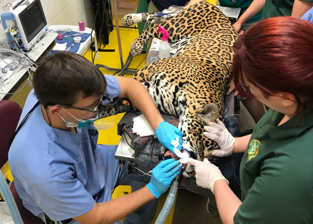 Dr Lewis performing the dental procedure.
Read our interview with Dr Lewis and an update on how Inka is doing in our next blog posts!