Better veterinary dental care through training
Blog post
26 March 2019
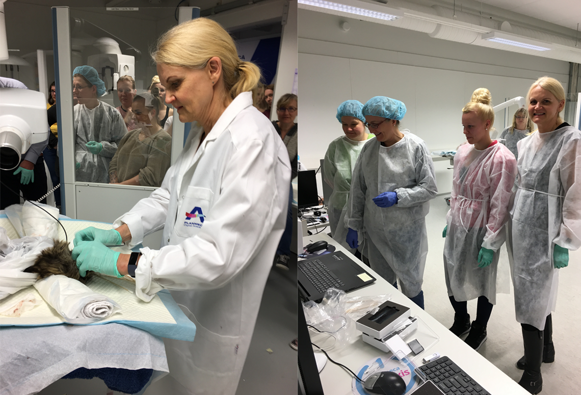
Dental specialist Eva Sarkiala shows how to take premium-quality intraoral X-ray images in a training for veterinarians.
As veterinary clinics are increasingly performing dental care, also the need for training is growing. Therefore, Planmeca Group is offering hands-on dental care education for veterinary professionals. Planmeca provides various trainings for veterinarians, from basic education to taking intraoral X-ray images and using the Planmeca Romexis Veterinary imaging software. Planmeca Group is proud to be cooperating with veterinarians who have extensive knowledge of dentistry and want to share their tips and tricks with their colleagues.
In the past years, the amount of dental care performed at the veterinary clinics has rapidly grown. However, since dentistry is not a big part of the veterinary education in most countries, training is often required when the veterinary clinics invest in dental equipment. The training at Planmeca includes both class room lectures and hands-on training for taking intraoral x-rays in the technical centre of Planmeca.
Planmeca offers versatile tools and services for veterinary professionals and has put a special emphasis on practical training. By acquiring practical experience, the veterinary professionals are able to provide the best dental care for animals. Planmeca provides various trainings for veterinarians, from basic education to taking intraoral X-ray images and using the Planmeca Romexis® imaging software as well as information about proper infection control.
As an example of veterinary training, Planmeca’s subsidiary in Finland, Plandent, teamed up with Evidensia_IVC veterinary group to offer their staff a full day of dental X-ray education. The training took place at the Planmeca training centre in Helsinki, with an access to high-quality intraoral X-ray devices and Planmeca Romexis veterinary software.

Intraoral X-ray images in the Planmeca Romexis® veterinary edition.
The participants had the opportunity to learn from the internationally renowned dental specialist Eva Sarkiala (DVM, DiplAVDC), as she shared her extensive knowledge on how to acquire premium-quality images. The vets had a chance to test both phosphor plates and sensors. The images acquired with the Planmeca ProSensor® HD intraoral sensor were viewed in the Planmeca Romexis software and showed a lot of detail. The participants also learnt how to process and interpret the X-ray images using the Planmeca Romexis imaging software for veterinary.
Text by: Mari Koivunen
Photos: Planmeca